Tuesday, December 16, 2014

Presepsin vs PCT

 2013 Jul 30;17(4):R168. doi: 10.1186/cc12847.

Diagnostic and prognostic value of presepsin in the management of sepsis in the emergency department: a multicenter prospective study.

Abstract

INTRODUCTION:

Sepsis, severe sepsis and septic shock are common conditions with high mortality. Their early diagnosis in the Emergency Department (ED) is one of the keys to improving survival. Procalcitonin (PCT) has been used as a biomarker in septic patients but has limited specificity and can be elevated in other scenarios of systemic inflammatory response syndrome (SIRS). Soluble CD14 (sCD14) or presepsin is the free fragment of a glycoprotein expressed on monocytes and macrophages. Preliminary reports suggest that levels of presepsin are significantly higher in septic patients than in healthy individuals. The aim of this study is to investigate the diagnostic and prognostic value of presepsin compared to PCT in people presenting at the ED with SIRS and suspected sepsis or septic shock.

METHODS:

This study was conducted in two major hospitals in Turin, Italy. One hundred six patients presenting to the EDs with suspected sepsis or septic shock were included, and another eighty-three patients affected by SIRS, but with no clinical evidence of infection, were recruited as controls. Blood samples were collected at first medical evaluation and for some patients after 24 and 72 h. The samples were analyzed using the PATHFAST Presepsin assay for sCD14, and commercial kits were used for other determinations (for example, PCT). Definitive diagnosis and survival rates were obtained afterward by analysis of digital medical records.

RESULTS:

Elevated concentrations of presepsin at presentation were observed in septic patients compared to control patients. The same trend was observed for mean values of PCT. Higher values of presepsin were observed in septic patients at presentation (time 0). The diagnostic accuracy of PCT was generally higher, and areas under the curve (AUCs) were 0.875 for PCT and 0.701 for presepsin. Mean presepsin values were significantly higher in nonsurvivor septic patients (60-day mortality) than in survivors. No significant correlation was noted between PCT and survival.

CONCLUSIONS:

In our experience, presepsin was useful in the early diagnosis of infection in a complex population of patients with SIRS, sepsis, severe sepsis and septic shock who presented to the ED. Presepsin showed a significant prognostic value, and initial values were significantly correlated with in-hospital mortality of patients affected by sepsis, severe sepsis or septic shock.

Massive vs Submassive PE

AHA PE Guidelines (Credit Emcrit)

Recently, the AHA published guidelines on the management of pulmonary embolism. I extracted only the stuff relevant to ED w/u and management:

Anticoagulation

Anticoagulate with LMWH, IV/Sub-Q UFH, or fondaparinux (IA)
While working up PE, if pretest is moderate or high, and there are no contra-indications, start anticoagulation during the work-up (IC)

Fibrinolytics

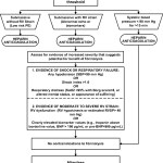
Definition of Massive PE-Acute PE with sustained hypotension (systolic blood pressure <90 mm Hg for at least 15 minutes or requiring inotropic support, not due to a cause other than PE, such as arrhythmia, hypovolemia, sepsis, or left ventricular [LV] dysfunction), pulselessness, or persistent profound bradycardia (heart rate <40 bpm with signs or symptoms of shock).
Definition of Submassive PE-Acute PE without systemic hypotension (systolic blood pressure >90 mm Hg) but with either RV dysfunction or myocardial necrosis.
  • RV dysfunction means the presence of at least 1 of the following:
    • —RV dilation (apical 4-chamber RV diameter divided by LV diameter >0.9) or RV systolic dysfunction on echocardiography
    • —RV dilation (4-chamber RV diameter divided by LV diameter >0.9) on CT
    • —Elevation of BNP (>90 pg/mL)
    • —Elevation of N-terminal pro-BNP (>500 pg/mL); or
    • —Electrocardiographic changes (new complete or incomplete right bundle-branch block, anteroseptal ST elevation or depression, or anteroseptal T-wave inversion)
  • Myocardial necrosis is defined as either of the following:
    • —Elevation of troponin I (>0.4 ng/mL) or
    • —Elevation of troponin T (>0.1 ng/mL)

Fibrinolysis is reasonable for pts with massive PE and acceptable risk of bleeding complications (IIa/B)
Fibrinolysis may be considered for pts with submassive PE judged to have clinical evidence of adverse prognosis (hemodynamic instability, worsening resp. insufficiency, severe RV dysfunction, or major myocardial necrosis) and low risk of bleeding complications (IIb/C)
Fibrinolysis is not recommended for patients with submassive PE with only mild dysfunction, i.e. low risk PEs (III/B)
Fibrinolysis is not recommended for undifferentiated cardiac arrest (III/B)
Interventional and Surgical Options

Either catheter embolectomy or surgical embolectomy can be considered depending on institutional and operator preference (IIa/C)
Either of these are reasonable if the pt is still unstable in massive PE after fibrinolysis (IIa/C)
Also reasonable in massive PE, if the pt has a contra-indication to lysis (IIa/C)
May be considered in lieu of fibrinolysis in patients with submassive PE and evidence of adverse prognosis (IIb/C)
Not recommended for pts with PE at low risk (III/C)

Contraindications to Fibrinolysis

Absolute contraindications include
  • any prior intracranial hemorrhage,
  • known structural intracranial cerebrovascular disease (eg, arteriovenous malformation),
  • known malignant intracranial neoplasm,
  • ischemic stroke within 3 months,
  • suspected aortic dissection,
  • active bleeding or bleeding diathesis,
  • recent surgery encroaching on the spinal canal or brain, and
  • recent significant closed-head or facial trauma with radiographic evidence of bony fracture or brain injury.
Relative contraindications include
  • age >75 years;
  • current use of anticoagulation;
  • pregnancy;
  • noncompressible vascular punctures;
  • traumatic or prolonged cardiopulmonary resuscitation (>10 minutes);
  • recent internal bleeding (within 2 to 4 weeks);
  • history of chronic, severe, and poorly controlled hypertension;
  • severe uncontrolled hypertension on presentation (systolic blood pressure >180 mm Hg or diastolic blood pressure >110 mm Hg);
  • dementia;
  • remote (>3 months) ischemic stroke; and
  • major surgery within 3 weeks.
Recent surgery, depending on the territory involved, and minor injuries, including minor head trauma due to syncope, are not necessarily barriers to fibrinolysis.
The clinician is in the best position to judge the relative merits of fibrinolysis on a case-by-case basis.

Further on who should get lytics

It is preferable to confirm the diagnosis of PE with imaging before fibrinolysis is initiated. When direct imaging is unavailable or unsafe because of the patient’s unstable condition, an alternative approach favors aggressive early management, including fibrinolysis, of the patient with sustained hypotension (systolic blood pressure <90 mm Hg for at least 15 minutes or requiring inotropic support, not clearly due to a cause other than PE) when there is a high clinical pretest probability of PE and RV dysfunction on bedside transthoracic echocardiography.We do not endorse the strategy of treating subjects with undifferentiated cardiac arrest with fibrinolysis, because this approach lacks clinical benefit.

PE Fibrinolytic Treatment Algorithm